Sun‑related skin changes often go unnoticed until they become a bigger problem. Spotting actinic keratosis at the first hint can keep you from facing a more serious diagnosis later on. This guide walks you through what the condition looks like, how to check your own skin, and when to call a professional.
Why catching it early matters
Actinic keratosis (AK) is a rough, scaly patch that forms on skin exposed to intense ultraviolet (UV) radiation. While many AKs stay harmless, a small percentage can evolve into squamous cell carcinoma, a type of skin cancer that can spread if ignored.
Detecting AK before it transforms gives you three clear advantages:
- Less invasive treatment options - topical creams or cryotherapy instead of surgery.
- Lower medical costs and fewer appointments.
- Peace of mind and reduced anxiety about skin health.
Understanding actinic keratosis
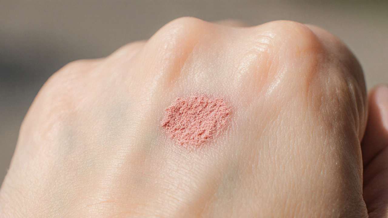
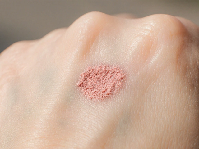
Actinic keratosis is a pre‑cancerous skin lesion caused by prolonged sun exposure. It typically appears on the face, ears, scalp, shoulders, neck, and back of the hands - any area that gets regular sunlight. The lesion feels gritty, like sandpaper, and may be tan, pink, red, or flesh‑colored.
Key attributes of AK:
- Size: usually 2‑10mm, though larger patches can develop.
- Texture: rough or crusty surface.
- Color: varies from light brown to deep red.
- Symptoms: often painless, but can itch or bleed if scraped.
Because AK looks similar to ordinary sun spots or even a harmless mole, learning the subtle differences is essential.
Spotting early signs: what to look for
The earliest AK lesions can be easy to miss. Keep an eye on these clues during a monthly self‑check:
- Texture change: A patch that feels rougher than the surrounding skin when you run a fingertip over it.
- Color shift: New pinkish or reddish tones that weren’t there before.
- Size growth: A spot that enlarges over weeks.
- Bleeding tendency: Minor bumps that bleed after light rubbing.
- Location pattern: Areas with high sun exposure - especially the dorsal hands and forehead.
If one or more of these signs appear, note the exact spot and date. Photographs taken in consistent lighting help track changes over time.
Step‑by‑step self‑examination
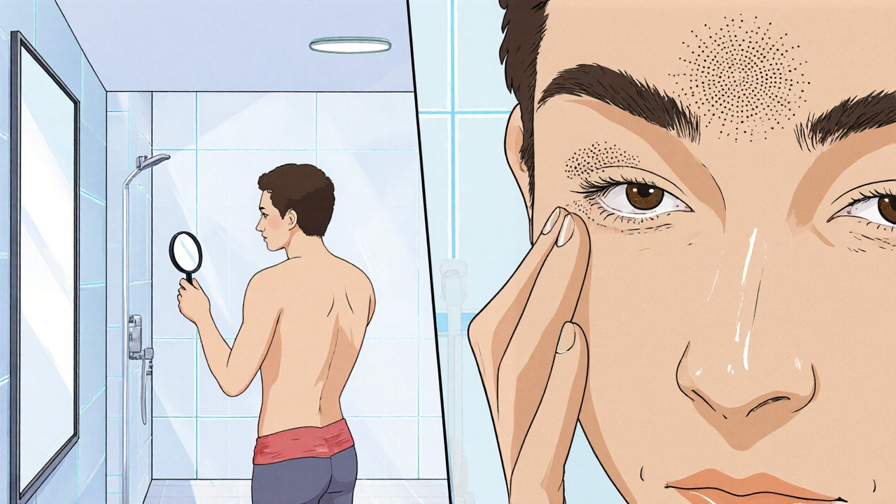
Set aside 10minutes in a well‑lit room, preferably after a shower when the skin is clean and softened. Follow these steps:
- Stand in front of a full‑length mirror and a handheld magnifying mirror.
- Start with your face. Look for any rough patches on the forehead, cheeks, nose, and ears.
- Move to the neck and shoulders, using the magnifier for hard‑to‑see spots.
- Raise your arms and examine the backs of your hands and forearms.
- Check the scalp if you have thinning hair - a hand‑held mirror can reveal hidden lesions.
- Feel each area gently; the gritty texture is a tell‑tale sign.
- Record any suspicious spots in a notebook or a secure phone app, noting size, color, and texture.
- If a lesion is new, changing, or bleeding, schedule a dermatologist appointment within two weeks.
Consistency is key: performing this routine once a month catches changes before they become problematic.
Risk factors and prevention tips
Knowing what raises your AK risk helps you stay ahead. Major risk factors include:
- Long‑term sun exposure - especially in regions with a high UV index.
- Fair skin, light hair, and a history of sunburns.
- Age over 40 - skin’s natural repair mechanisms weaken.
- Immunosuppression, such as after organ transplantation.
- Certain medications that increase photosensitivity.
Preventive actions that lower AK chances:
- Apply broad‑spectrum sunscreen with SPF30 or higher every day, even when it’s cloudy.
- Wear protective clothing - wide‑brimmed hats, long sleeves, and UV‑blocking sunglasses.
- Seek shade between 10am and 4pm when UV intensity peaks.
- Consider regular professional skin checks, especially if you’ve had AK before.
When to see a dermatologist
A dermatologist is a skin specialist who can confirm an AK diagnosis and recommend treatment. Schedule an appointment if:
- Any lesion fits the early‑sign checklist and persists for more than two weeks.
- You notice bleeding, crusting, or an ulcerated spot.
- There are multiple lesions in a concentrated area.
- You have a personal or family history of skin cancer.
During the visit, the doctor may perform a dermatoscopic exam, shave biopsy, or cryotherapy, depending on the lesion’s appearance.

Comparing common sun‑related lesions
| Feature | Actinic Keratosis | Solar Lentigo (Age Spot) | Basal Cell Carcinoma | Melanoma |
|---|---|---|---|---|
| Typical color | Pink, red, or brown; may be flesh‑colored | Uniform brown or black | Translucent, pinkish pearly | Varied - black, brown, blue, red |
| Texture | Rough or sandpaper‑like | Smooth | Shiny, may ulcerate | Often uneven, may have itching or bleeding |
| Common locations | Face, forearms, scalp, hands | Hands, forearms, shoulders | Nose, ears, cheeks | Any body part, often back, legs |
| Potential to become cancer | Low‑to‑moderate (can turn into SCC) | None | Low‑grade skin cancer (local growth) | High‑grade, can metastasize |
| Treatment urgency | Prompt removal recommended | Cosmetic only | Early excision advised | Immediate specialist care |
Understanding these distinctions helps you decide whether a lesion warrants a quick doctor’s visit or can be monitored at home.
Quick self‑check checklist
- Do you have any rough, sandpaper‑like spots on sun‑exposed skin?
- Are any of those spots red, pink, or different in color from surrounding skin?
- Has any spot grown in size over the past few weeks?
- Do any lesions bleed after light rubbing?
- Is your sun protection routine adequate for your skin type?
If you answered “yes” to two or more, it’s time to document the spots and book a dermatologist appointment.
Frequently Asked Questions
Can actinic keratosis go away on its own?
Occasionally a small AK may flatten, but most persist and can turn into cancer if untreated. Professional removal is the safest option.
How often should I perform a self‑examination?
A monthly routine catches new lesions early. Increase frequency if you notice any changes between check‑ups.
What treatments are available for early‑stage AK?
Topical creams (5‑fluorouracil, imiquimod), cryotherapy with liquid nitrogen, photodynamic therapy, and surgical excision are common choices. Your dermatologist will recommend what fits your lesion count and skin type.
Is sunscreen enough to prevent actinic keratosis?
Sunscreen greatly reduces risk, but it’s not a guarantee. Combining sunscreen with protective clothing and limiting peak‑hour sun exposure provides the best defense.
Should I be worried if I have a family history of skin cancer?
A family history raises your baseline risk, making regular self‑checks and professional skin exams even more important.

John Chapman
October 14, 2025 at 20:46While the piece does a decent job outlining the visual cues of actinic keratosis, it omits the dermoscopic patterns that can decisively differentiate AK from benign solar lentigines. In practice, the presence of a ‘strawberry pattern’ and rosette structures under polarized light are highly predictive. Moreover, the guide fails to address the role of field cancerisation, which underlies the multiplicity of lesions in high‑risk patients. For clinicians, recommending a full‑body digital dermoscopy set‑up can dramatically improve early detection rates. Finally, educating patients about the incremental risk per lesion-approximately 0.1% progression to SCC-is essential for informed consent.
Tiarna Mitchell-Heath
October 24, 2025 at 03:00Are we really supposed to swallow this cookie‑cutter checklist and call it medical advice? It reads like a repackaged brochure from a sunscreen manufacturer and lacks any real nuance.
Katie Jenkins
November 2, 2025 at 08:13First, the definition of actinic keratosis presented at the top is accurate, but the article should clarify that not all rough, scaly patches are AK-some are merely seborrheic keratoses or lichen planus. Second, the recommended monthly self‑examination frequency is reasonable, yet many dermatologists advise a bi‑weekly check during peak UV months for patients with a history of skin cancer. Third, the texture description ("sandpaper‑like") is useful, but adding that the lesion often feels firm rather than floppy can help novices. Fourth, the color spectrum noted (pink, red, brown) is comprehensive, but the guide neglects the occasional yellowish hue seen in hyperkeratotic AK. Fifth, the guide’s emphasis on photography is spot‑on; however, using a consistent neutral background prevents false color perception. Sixth, the step‑by‑step list correctly includes a magnifying mirror, but it should also mention a dermatoscope for clinicians. Seventh, the advice to record lesions in a notebook is solid, yet a digital app with measurement tools provides more precise tracking. Eighth, the article states that "most AKs stay harmless," which is technically true, but the 0.5% to 1% malignant transformation rate is not negligible. Ninth, the risk factor section accurately cites fair skin and age, but it omits immunosuppressive medications like azathioprine. Tenth, sunscreen usage is highlighted, but the guide fails to warn that re‑application every two hours is mandatory for true protection. Eleventh, the table comparing lesions is helpful, yet it could benefit from a column on typical dermoscopic features. Twelfth, the "when to see a dermatologist" checklist is clear, but adding a note about teledermatology options would modernize it. Thirteenth, the treatment options overview is concise, yet it should differentiate between lesion‑directed therapy (cryotherapy) and field‑directed therapy (5‑fluorouracil). Fourteenth, the FAQ about self‑resolution accurately notes that spontaneous regression is rare, but references to clinical studies would bolster credibility. Fifteenth, the article encourages protective clothing, but including UPF‑rated fabrics would be more specific. Finally, overall the guide is a solid foundation for laypeople, but integrating the above refinements would elevate it from good to exemplary.
Roger Wing
November 11, 2025 at 14:26Don't be fooled-most of what you read about "protective sunscreen" is a marketing ploy pushed by big pharma to sell more lotions. The real protection comes from staying indoors during peak UV hours and wearing a hat, not from a bottle of chemicals.
Alison Poteracke
November 20, 2025 at 20:40Great job breaking down the steps! I’ve started using the monthly mirror routine and it’s already helped me spot a tiny patch before it got bigger. Keep the tips coming, they’re super helpful.
Marianne Wilson
November 30, 2025 at 02:53While enthusiasm is commendable, the guide glosses over the fact that many readers will skip the "raise your arms" step, missing lesions on the dorsal hands where AKs are most prevalent. A more forceful reminder would improve compliance.
Patricia Bokern
December 9, 2025 at 09:06Whoa, this guide feels like a drama series where every episode ends with a new skin monster. I swear my own elbows look like they’re auditioning for a horror film after a week in the sun.
Garrett Gonzales
December 18, 2025 at 15:20In clinical terms, cryotherapy with liquid nitrogen achieves a rapid freeze-thaw cycle that destroys atypical keratinocytes within minutes. The optimal spray duration is 10–15 seconds per lesion, followed by a brief observation period for erythema. For thicker AKs, a double-freeze technique-30 seconds, thaw, then another 30 seconds-has shown higher clearance rates. Post-treatment care includes a bland moisturizer and sun avoidance for at least 48 hours to minimize post-inflammatory hyperpigmentation.
Aman Deep
December 27, 2025 at 21:33🌞 Life under the sun is a fleeting canvas, and every speck of sand-paper skin tells a story of our fleeting choices. 🌱 If we paint with shade and gratitude, the canvas stays vibrant. 😌 Remember, the skin is the map of our journeys; walk wisely. 🌟
Chris Smith
January 6, 2026 at 03:46Oh wow, another “must-do” list. Because we all have time to stare at our hands all day.
Camille Ramsey
January 15, 2026 at 10:00Listen, the guide is riddled with sloppy phrasing-"often painless" should be "generally painless," and "may itch" is vague. Also, "apply sunscreen with SPF30" is a minimum; dermatologists recommend SPF50 for high-risk patients. Lastly, the phrase "once a month" ignores the fact that lesions can change in days, not weeks. Fix these and you’ll sound professional.
Raghav Narayan
January 24, 2026 at 16:13It is understandable that many individuals feel overwhelmed by the prospect of regular self-examinations, yet the psychological benefits of empowerment should not be underestimated. By allocating merely ten minutes in a well-lit environment, one cultivates a habit that seamlessly integrates into routine personal care. The systematic approach-starting at the face, progressing to the neck and shoulders, then the extremities-mirrors the anatomical gradient of sun exposure, thereby maximizing detection efficiency. Furthermore, documenting each observation in a secure digital journal not only creates a chronological record but also facilitates more accurate communication with dermatology professionals, should a referral become necessary. In addition, reinforcing sun-protective behaviors, such as wearing UPF-rated garments and seeking shade during peak ultraviolet hours, complements the self-examination protocol and helps to reduce the incidence of new lesions. Ultimately, the combination of vigilant inspection, meticulous documentation, and proactive photoprotection forms a comprehensive strategy that significantly diminishes the likelihood of actinic keratosis progressing to squamous cell carcinoma.
Tara Phillips
February 2, 2026 at 22:26Esteemed community, I commend the author for an exhaustive and methodical exposition. May your diligent self-checks inspire countless individuals to uphold their cutaneous health with unwavering dedication. Let us all pursue preventative vigilance with steadfast resolve.
Derrick Blount
February 12, 2026 at 04:40In regard to the presented checklist, one must observe, that the inclusion of "photographs in consistent lighting" is indispensable; without it, longitudinal comparison becomes, frankly, untenable. Moreover, the recommendation to "schedule a dermatologist appointment within two weeks" should, arguably, be contingent upon lesion size, as larger lesions merit more immediate consultation. Lastly, the suggestion to "apply broad-spectrum sunscreen" ought to be expanded to encompass re-application intervals, which, by consensus, are every two hours during sun exposure.
Priya Vadivel
February 21, 2026 at 10:53The emphasis on supportive community sharing is heartening; it reminds us that skin health is not solely a personal battle but a collective journey. By encouraging others to document their observations, we foster a culture of accountability and early intervention. Such solidarity can reduce the emotional burden often associated with dermatological concerns.
Dharmraj Kevat
March 2, 2026 at 17:06Drama aside, the truth is simple: protect your skin or pay the price later.
Lindy Fujimoto
March 11, 2026 at 23:20Honestly, this guide is like a runway show for sun-savvy fashion! 🌞✨ Your tips are the glitter that makes every skin-care routine sparkle. Keep dazzling us with that brilliance, and don’t forget to add a splash of emoji flair! 💖👑
darren coen
March 21, 2026 at 05:33Well done, very useful.